Robotic microscope used for first time in pioneering brain surgery
A neurosurgical team at The Royal London Hospital has for the first time in Europe used an innovative digital robotic microscope to successfully operate on a dangerous brain aneurysm (bulging blood vessel), potentially reducing the length of such operations.
With a state-of-the-art digital microscope positioned on the end of a robotic arm, the device - called a Modus V - is a completely hands free digital robotic microscope. It uses infra-red technology to follow the surgeons’ movements during operations, automatically changing its position, effectively delivering light and automatically focusing on the tip of the instrument. Critically, this means that the brain is always in focus throughout the operation. It was made possible using a £500K grant from Barts Charity.
The new device is safer for patients by allowing the surgeon to remain in a comfortable and steady position throughout the operation while the robotic microscope follows their movements. With conventional microscopes, every time the microscope moves the surgeon also has to move, effectively following the microscope. Changing the microscope's position interrupts the flow of the operation, increasing the already lengthy operation by up to 10 per cent.
Current (optical) microscopes are also limited in the positions that they can be put into since the surgeon has to be able to look directly into the eye pieces while being comfortable enough to steadily perform microsurgery.
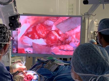
The image is projected onto a 55 inch high definition screen, vastly improving the view for other members of the team.
The Royal London Hospital Neurosurgeon Chris Uff was the first in Europe to successfully use the device to repair a brain aneurysm.
Chris explained: “We are now able to operate in a completely new and improved way. Everyone in the operating theatre has the same view that I do, so my assistant and the nurses know exactly what my next move will be. If there is a risk we can all react immediately. I don't have to change my position or the patient's position and I can put the microscope anywhere I choose - in this operation I saw angles of approach I have never seen before. The improved image quality is like the difference between using binoculars and watching a wide-screen HD TV.”
Mr Uff was the first surgeon in Europe to use the exoscope to repair a brain aneurysm on 57 year old Hasibe Behjet. After three days’ recovery at The Royal London Hospital she was able to return home to Hackney.
Hasibe said: “It’s wonderful to be the first person to benefit from this new equipment. Mr Uff and his team are really lovely and explained everything so clearly."
Small brain aneurysms are common, occurring in about 1 in 30 people. While the majority are harmless, larger aneurysms are more likely to bleed which can be deadly.
Fiona Miller Smith, Barts Charity Chief Executive added: "Equipment like this and the Da Vinci surgical robots that we funded in 2017, keeps Barts Health NHS Trust at the forefront of digital technology in surgery. We’re delighted that the exoscope is already improving the surgical experience for brain surgeons and their patients."